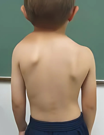
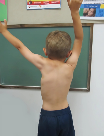
Torticollis, sprengel shoulder and other scapular deformity
What is congenital muscular torticollis?
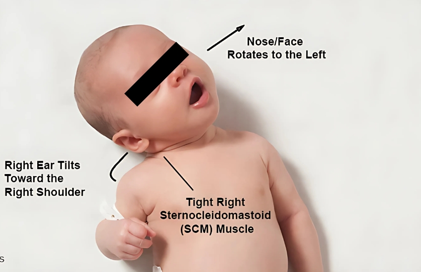
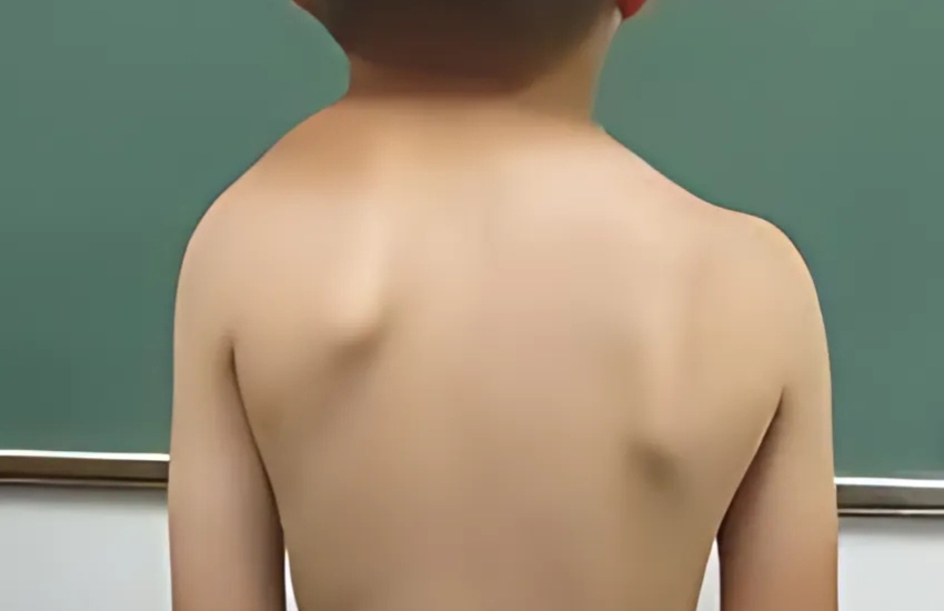
Congenital muscular torticollis is a condition in which an infant’s neck muscle is shortened causing the neck to twist. Congenital means present at birth and torticollis means twisted neck. The condition is sometimes called wryneck.
Causes
Congenital muscular torticollis may occur following a difficult birth, especially if the infant is delivered breech. During the delivery, if the sternocleidomastoid muscle, the neck muscle that extends from the jawbone (mastoid) to the clavicle (collarbone) and sternum (breastbone), is stretched or pulled, it may tear, causing bleeding and bruising within the muscle. The injured muscle develops fibrosis (scar tissue) which causes the muscle to shorten and tighten, pulling the infant’s head to one side. The fibrosis forms a mass or lump that sometimes can be felt on the side of the neck.

Symptoms
Congenital muscular torticollis may be visible at birth or it may not become evident until several weeks later. The following are the most common symptoms of congenital muscular torticollis. However, each child may experience symptoms differently. Symptoms may include:
-
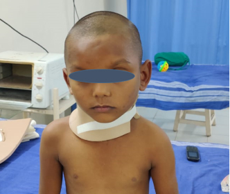
Tilting of the infant’s head to one side
-
The infant’s chin turns toward the opposite side of the head
-
Firm, small, one to two centimeter mass in the middle of the sternocleidomastoid muscle
The symptoms of congenital muscular torticollis may resemble other neck masses or medical problems. Always consult your child’s doctor for a diagnosis.
Diagnosed
Generally, physical examination of the infant may show the characteristic tilting of the head and tension of the sternocleidomastoid muscle, as well as presence of a mass in the middle portion of the muscle. In addition to a complete medical history and physical examination, diagnostic procedures for congenital muscular torticollis may include the following:
-
X-rays. A diagnostic test that uses invisible electromagnetic energy beams to produce images of internal tissues, bones, and organs onto film to check for abnormalities in the bones of the neck and shoulders.
-
Ultrasound examination. A diagnostic imaging technique that uses high-frequency sound waves and a computer to create images of blood vessels, tissues, and organs to evaluate the muscle around the mass. Ultrasounds are used to view internal organs as they function and to assess blood flow through various vessels.
Treatment
If the condition is not corrected, the infant will be unable to move his or her head properly. Permanent muscle tightening with asymmetry (uneven development) of the neck and face can result. Specific treatment of congenital muscular torticollis will be determined by your child’s doctor based on:
-
Your child’s age, overall health, and medical history
-
Extent of the condition
-
Your child’s tolerance for specific medications, procedures or therapies
-
Expectations for the course of the condition
-
Your opinion or preference
Treatment may include:
-
Gentle stretching exercise program (to help relieve the tension and lengthen the sternocleidomastoid muscle)
-
Infant stimulation (to help the infant learn to move and stretch the muscle)
-
Surgery (to correct the shortened muscle)
Pre-OP & Post-OP Pictures
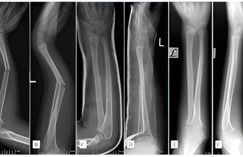
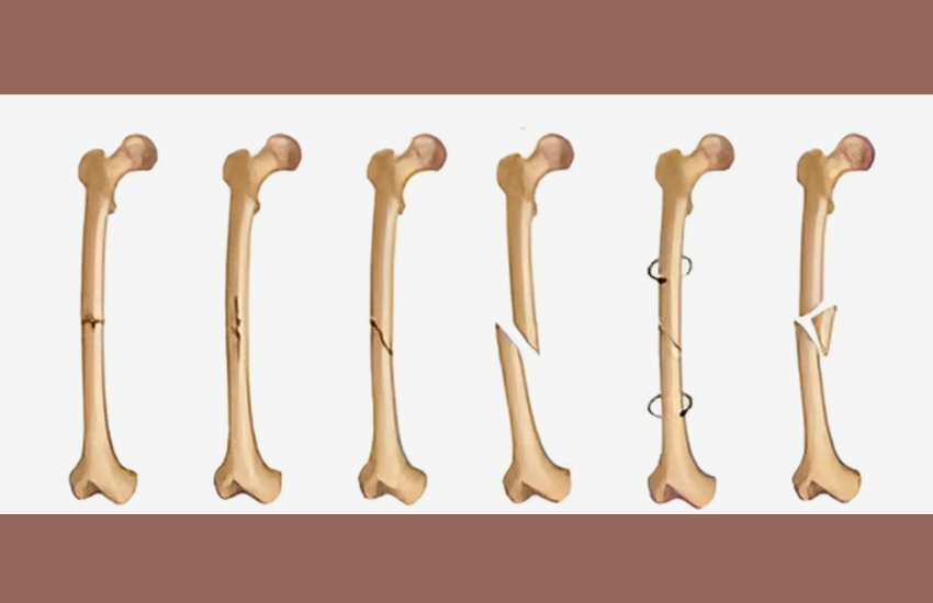
Post trauma (Exposed bone)
Paediatric forearm Open fracture with exposed bone
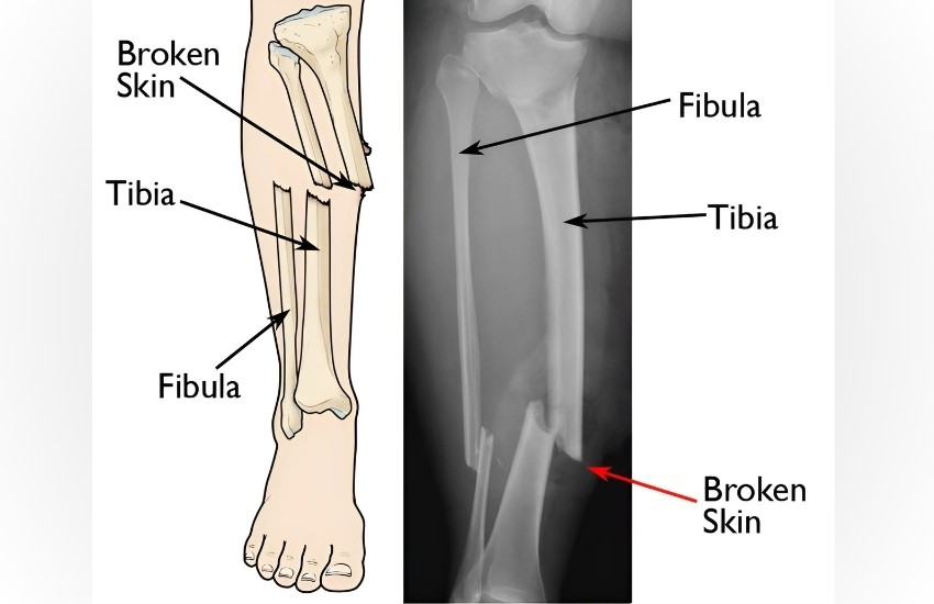
Road Traffic Accident with open wound
Road Traffic Accident with Open tibia fracture advised amputation elsewhere saved his leg with multiple surgery
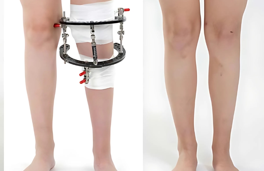
Bone Lengthening
Gradual Bone Lengthening using Orthofix
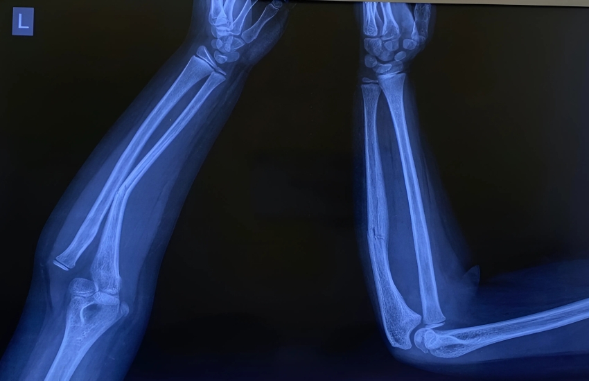
MONTEGGIA FRACTURE DISLOCATION
Monteggia Fracture dislocation treated with plaster without surgery
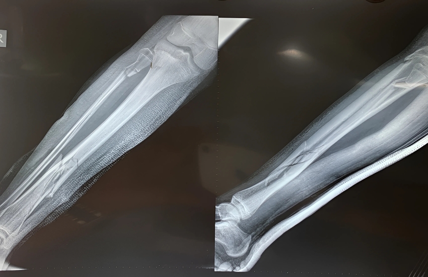
Open Tibia Fracture
Open Tibia Fracture managed with elastic nail and JESS fixator
Bone and soft tissue tumors
Minimally invasive surgery to correct the deformity without cutting the bone with single screw (day care surgery)
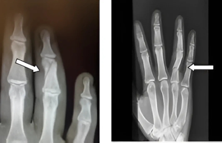
Middle Phalanx Fracture
Neglected Middle Phalanx Fracture managed with minimally invasive surgery with mini JESS Fixator
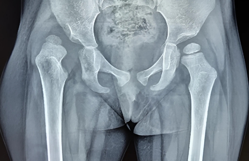
DDH xray
3 years old child with right hip Dysplasia with dislocation underwent open reduction and femoral shortening and varus derotation osteotomy
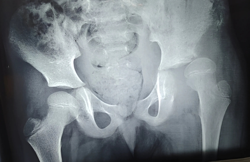
Hip Joint
4 years old girl with left hip joint subluxation with coxa valga (post septic) underwent left hip Open Reduction and proximal femur varus derotation osteotomy and hip spica
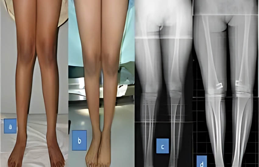
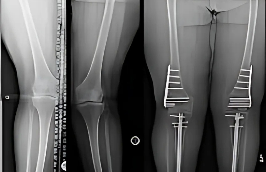
GENU VALGUM PREOP AND POST OP IMAGES
Bilateral genu valgus deformity correction with osteotomy
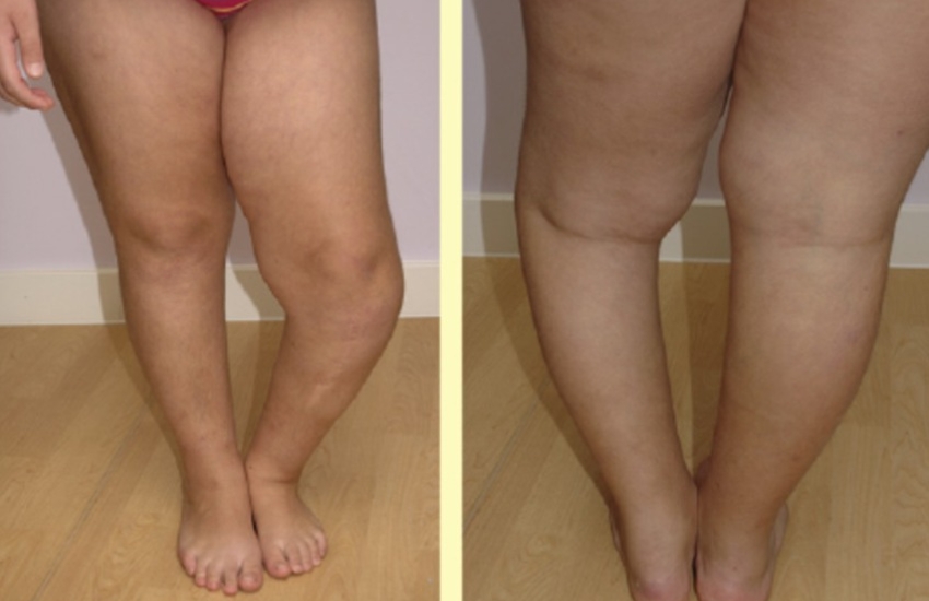
Deformity
knee deformity (blount disease) genu varum left side
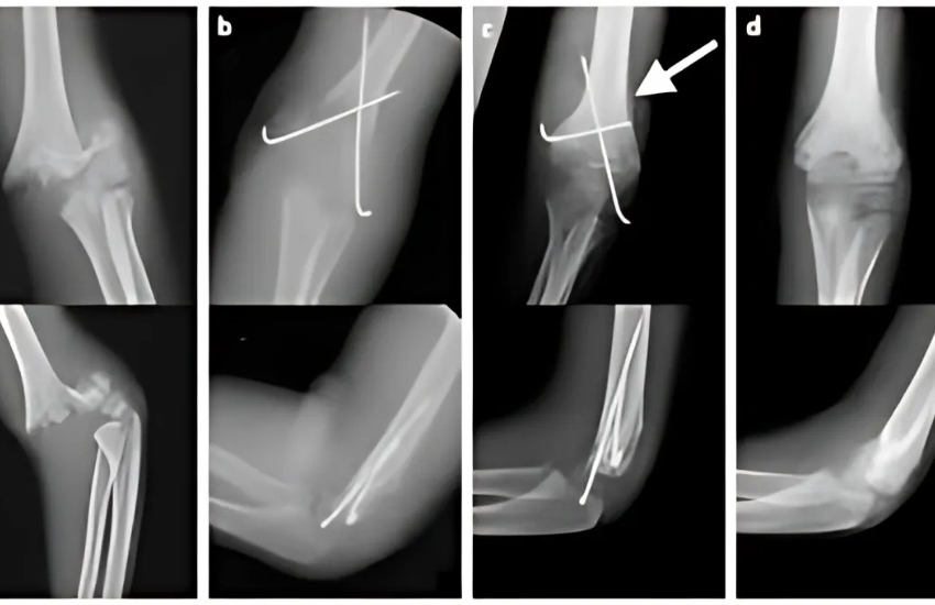
Supracondylar Fracture
This is supracondylar fracture in a 5yr old child after slip and fall while playing ( x ray before surgery )
SC Fixation
SC# AP view after reduction and fixation
Road Traffic Accident
AFTER ankle and femur fixation surgery